- Introduction
- About Your Breasts
- Understanding Cancer
- Risk Factors
- Screening
- Symptoms
- Diagnosis
- Additional Tests
- Staging
- Treatment
- Breast Reconstruction
- Complementary and Alternative
Medicine - Nutrition and Physical Activity
- Follow-up Care
- Sources of Support
- The Promise of Cancer Research
- National Cancer Institute Information
Resources
Introduction
This United Cancer Foundation has important information about breast cancer. Breast cancer is the most common type of cancer among women in this country (other than skin cancer). Estimates for 2008 are that more than 182,000 American women learn they have this disease.
You will read about possible causes, screening, symptoms, diagnosis, treatment, and supportive care. You will also find ideas about how to cope with the disease.
Scientists are studying breast cancer to find out more about its causes. And they are looking for better ways to prevent, find, and treat it.
The Breasts
The breasts sit on the chest muscles that cover the ribs. Each breast is made of 15 to 20 lobes. Lobes contain many smaller lobules. Lobules contain groups of tiny glands that can produce milk. Milk flows from the lobules through thin tubes called ducts to the nipple. The nipple is in the center of a dark area of skin called the areola. Fat fills the spaces between the lobules and ducts.
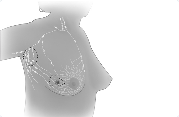
The breasts also contain lymph vessels. These vessels lead to small, round organs called lymph nodes. Groups of lymph nodes are near the breast in the axilla (underarm), above the collarbone, in the chest behind the breastbone, and in many other parts of the body. The lymph nodes trap bacteria, cancer cells, or other harmful substances.
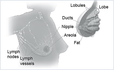
These pictures show the parts of the breast and the lymph nodes and lymph vessels near the breast.
About Your Breasts
Breast Basics
The breast is a gland that produces milk in late pregnancy and after childbirth.
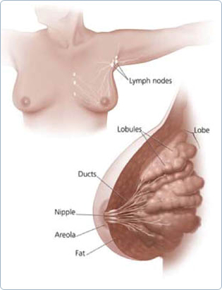
What are breasts made of?
- Each breast is made of lobes.
- Lobes are groups of milk glands called lobules.
- Lobules are arranged around thin tubes called ducts.
- Ducts carry the milk to the nipple.
- These lobules and ducts make up the glandular tissue.
What is the lymphatic system?
The breasts also contain lymph vessels, which carry a clear fluid called lymph.
» The lymph vessels lead to small, round organs called lymph nodes. Groups of lymph nodes are found near the breast in the underarm, above the collarbone, in the chest behind the breastbone, and in many other parts of the body.
» The lymph nodes trap bacteria, cancer cells, or other harmful substances that may be in the lymphatic system. Their job is to make sure harmful substances are safely removed from the body.
See your health care provider about a breast change when you have:
- A lump in or near your breast or under your arm
- Thick or firm tissue in or near your breast or under your arm
- Nipple discharge or tenderness
- nipple pulled back (inverted) into the breast
- Itching or skin changes such as redness, scales, dimples, or puckers
- A change in breast size or shape
If you notice a lump in one breast, check the other breast. If both breasts feel the same, it may be normal. You should still see your health care provider for a clinical breast exam to see if more tests are needed.
Types of Breast Changes
Breast changes occur in almost all women. Most of these changes are not cancer. However, some breast changes may be signs of cancer. Breast changes that are not cancer are called benign. For more information on benign breast changes that are more likely to happen during certain times of your life, see Breast Changes During Your Lifetime That Are Not Cancer.
Lumpiness
Most women have some type of lumpiness in their breasts. Some areas may be more dense than others and can feel lumpy in an exam. What you are feeling may be glandular breast tissue.
Breast Changes Due to Your Period
Many women have swelling, tenderness, and pain in their breasts before and sometimes during their periods. You may also feel one or more lumps during this time because of extra fluid in your breasts.
Because some lumps are caused by normal hormone changes, your health care provider may suggest watching the lump for a month or two to see if it changes or goes away.
Single Lumps
Single lumps can appear at any time and come in various types and sizes. Most lumps are not cancer, but your health care provider should always check the lump carefully. He or she may do more tests to make sure the lump is not cancer. See Chart 1: Possible Mammogram Results and Follow-Up Care for more information about these types of lumps.
Check with your health care provider if you notice any kind of lump. Even if you had a lump in the past that turned out to be benign, you can't be sure that a new lump is also benign.
Nipple Discharge
Nipple discharge is common for some women. It is fluid that comes from the nipple in different colors or textures. Usually, it is not a sign of cancer. For example, birth control pills and other medicines, such as sedatives, can cause a little discharge. Certain infections also cause nipple discharge. However, for women who are going through or have passed menopause, nipple discharge can be a sign of cancer.
See your doctor if you have nipple discharge for the first time, or a change in your discharge's color or texture. He or she may send a sample of the discharge to be checked at a lab.
Finding Breast Changes
There are two ways to find breast changes:
- Clinical breast exams--a breast exam done by your health care provider
- Mammograms --an x-ray of your breasts
One way to find breast changes is with a clinical breast exam done by your health care provider. He or she will check your breasts and underarms for any lumps, nipple discharge, or other possible changes. This breast exam should be part of a routine medical check-up.
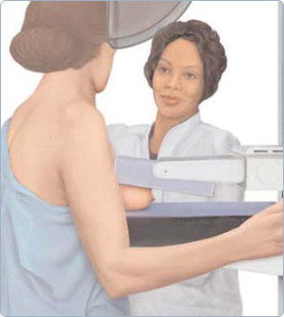
The best tool for finding breast cancer is a mammogram. A mammogram is a picture of the breast that is made by using low-dose x-rays. It is currently recommended that women over age 40 receive a mammogram every 1 to 2 years.
Some women check their own breasts for changes. If you find a change, it's important to call your health care provider for an appointment. Make sure to watch the change you found until you see your provider. But a breast self-exam and a clinical breast exam are not substitutes for mammograms.
Questions to ask your health care provider about a breast change
- How can I tell the difference between my usual lumps and lumps I need to do something about?
- How will you be able to tell what kind of breast change I have?
- What should we do to watch this change over time?
Mammograms
Mammograms are used for both screening and diagnosis.
A screening mammogram is used to find breast changes in women who have no signs of breast cancer. Most women get two x-rays of each breast.
If your recent screening mammogram revealed a breast change since your last one, or if you or your health care provider noticed a change, he or she will probably recommend a diagnostic mammogram. More x-rays are taken during a diagnostic mammogram than a screening mammogram to get clearer, more detailed pictures of the breast. It is also used to rule out other breast problems.
Tip
Take your original mammogram and copy of the medical report with you if you change doctors or centers or need a second opinion.
A digital mammogram is another way to take a picture of your breasts. The procedure for having a digital mammogram is the same as a screening mammogram, except that it records the x-ray images in computer code instead of on x-ray film.
It is important to see your doctor and get a mammogram every 1 to 2 years after age 40 to find breast changes.
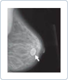
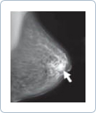
Here are mammograms of three different women. Doctors look at these x-rays for any breast changes that don't look normal.
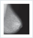
(Not Cancer)
Mammograms and Breast Implants
When you go for your mammogram, tell staff if you have a breast implant. A technologist who is trained in x-raying patients with implants will do your mammogram. Breast implants can hide some breast tissue and make it harder to read your mammograms.
If you got implants for cosmetic reasons:
- You still need to get screening mammograms, with extra pictures to help get an accurate reading.
If you got an implant after having a mastectomy for breast cancer:
- You should continue to get mammograms of your other breast. Ask your doctor if you still need mammograms of the breast with the implant.
Getting Your Mammogram Results
Ask your doctor when you will get your results. You should get a written report of your mammogram results within 30 days of getting the x-ray. This is the law. Be sure the mammogram facility has your current address.
If your results were normal, it means the radiologist did not find anything that needs follow-up.
If your results were abnormal, it means the radiologist found:
- A change from a past mammogram
- A change that needs more follow-up
What a Mammogram Can Show
The radiologist will look at your x-rays for breast changes that do not look normal. The doctor will look for differences between your breasts. He or she will compare your past mammograms with your most recent one to check for changes. The doctor will also look for lumps and calcifications.
Lumps (or "mass")
The size, shape, and edges of a lump sometimes can give doctors more information about whether or not it is cancer. On a mammogram, a growth that is benign often looks smooth and round with a clear, defined edge. On the other hand, breast cancer often has a jagged outline and an irregular shape. (See Chart 1: Possible Mammogram Results and Follow-Up Care.)
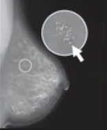
Calcifications
A calcification is a deposit of the mineral calcium in the breast tissue. Calcifications appear as small white spots on a mammogram. There are two types:
- Macrocalcifications are large calcium deposits often caused by aging. These are usually not cancer. (See Chart 1: Possible Mammogram Results and Follow-Up Care.)
- Microcalcifications are tiny specks of calcium that may be found in an area of rapidly dividing cells. If they are found grouped together in a certain way, it may be a sign of cancer. (See Chart 1: Possible Mammogram Results and Follow-Up Care.)
Depending on how many calcium specks you have, how big they are, and what they look like, your doctor may suggest that you:
- Have a different type of mammogram that allows the radiologist to have a closer look at the area
- Have another screening mammogram, usually within 6 months
- Have a test called a biopsy.

Calcium in the diet does not create calcium deposits (calcifications) in the breast.
Calcification

Anne, Age 60
Are Mammogram Results Always Right?
No. Although they are not perfect, mammograms are the best method to find breast changes. If your mammogram shows a breast change, sometimes other tests are needed to better understand it. Even if the doctor sees something on the mammogram, it does not mean it is cancer.
Changes That Need More Follow-Up
Sometimes your doctor needs more information about a change on your mammogram. Your doctor may do follow-up tests such as an ultrasound or more mammograms. The only way to find out if an abnormal result is cancer is to do a biopsy. It is important to know that most abnormal findings are not cancer.
Breast Changes During Your Lifetime That Are Not Cancer
You might notice different kinds of breast changes at different times in your life. Many of these are caused by changes in your hormone levels and are a normal part of getting older.
Younger women may have more glandular (more dense, less fatty) breast tissue than older women who have stopped having their period (menopause). This kind of tissue is where breast changes usually occur.
Before or during your period, you might have lumpiness, tenderness, and pain in your breasts. The lumpiness and pain usually go away by the end of your period.
During pregnancy, your breasts may feel lumpy, as the glands that produce milk increase in number and get larger. Still, breast cancer has been found in pregnant women, so talk with your doctor if you have questions about any breast lumps.
Carmen, Age 70
While breastfeeding, you may get an infection called mastitis that happens when a milk duct becomes blocked. Mastitis causes the breast to look red and feel lumpy, warm, and tender. Mastitis is often treated with antibiotics. Sometimes the duct may need to be drained. If the redness or mastitis does not go away with treatment, call your doctor, as you may need further care.
As you approach menopause, your periods may become less frequent. Changing hormone levels also can make your breasts:
- Feel tender, even when you are not having your period
- Feel more dense
- Feel more "lumpy" than they did before
As you age, other breast changes are more common, such as:
- Intraductal papilloma. This is a growth inside the nipple that looks like a wart. It can be removed by surgery without changing the way the breast looks.
- Mammary duct ectasia. As you near menopause, ducts beneath the nipple can become swollen and clogged. This can be painful and cause nipple discharge. The problem is treated with warm packs, antibiotics, and sometimes surgery to remove the duct.
If you are taking hormones, such as hormone replacement therapy (HRT), birth control pills, or injections, when getting your mammogram be sure to let your doctor know. Hormones may cause your breasts to be more dense. This can limit your doctor's ability to read a mammogram.
When you stop having periods (menopause), your hormone levels drop, and your breast tissue becomes less dense and more fatty. You may stop having the lumps, pain, or nipple discharge you used to have. And because your breast tissue is less dense, mammograms can be easier to read. This means doctors are more likely to find breast changes or early breast cancer.
Getting a Second Opinion
Marie, Age 55
After seeing your health care provider, you may want to get a second opinion from another health care provider. Sometimes, a second opinion may be covered by your health insurance. Many women feel uneasy about asking for a second opinion, but it is very common. Your health care provider will not be surprised or offended if you seek a second opinion. A second opinion might help you feel more confident that you are making the best choices about your health.
Follow-Up Tests to Tell You More
Doctors often use ultrasound or biopsies to follow up after finding signs of a breast change.
Ultrasound
An ultrasound uses sound waves to make a picture of breast tissue. This picture is called a sonogram. It helps doctors look more closely at lumps. An ultrasound shows if a lump is solid or filled with fluid (cyst). (See Chart 1: Possible Cysts are usually not cancer. Mammogram Results and Follow-Up Care.) An ultrasound also can help your doctor decide if more tests are needed.
It is important to know that an ultrasound may not find all abnormal changes.

A Cyst: The Most Common Breast Change
A cyst is a lump filled with fluid.
If a cyst is found, your doctor may decide to:
- Remove fluid in the cyst to make it go away
- Look at it with an ultrasound
- Watch it closely over time

Biopsies
During a biopsy, the doctor removes some cells or tissue, an area of small calcium deposits, or the whole lump. The tissue is sent to a lab where a doctor called a pathologist will look at the cells. He or she checks the cells for cancer or other diseases. Biopsies are usually done on an outpatient basis, meaning you can go home the same day as your test. Biopsies are the only way to find out if cells are cancer.
Questions you might want to ask if your health care provider suggests a biopsy:
- What type of biopsy will I have? Why? How much tissue will be removed?
- Will only part of the lump or the whole lump be removed?
- Will the shape of my breast change?
- Will I have a scar?
- Where will the biopsy be done? How long will it take?
- Will I be awake?
- Will it hurt?
- Will there be any side effects?
- What tests will be done on the tissue?
- When will I know the results?
- Will I be able to go back to work the same day?
After the biopsy, you might want to ask:
- What do the results mean?
- Do I need to have other tests?
- Will I need follow-up tests or treatment?
- How do I care for the biopsy site?
- Is it okay if I exercise?
- Does my result mean I am at higher risk for breast cancer?
Sometimes it's hard--even for the experts--to tell benign breast changes from cancer. If you have any questions about your biopsy results, have more than one doctor look at the results.
Types of Biopsies
The most common types of biopsies are:
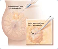
Fine-Needle Aspiration
Fine-needle aspiration only takes a few minutes and can be done in the doctor's office. It is often done when a doctor finds a lump that appears to be a cyst. The doctor tries to remove fluid from the lump using a thin needle and a syringe. If the lump is a cyst, removing the fluid will make it go away. If the cyst returns, it can be drained again. Because cysts are rarely cancer, doctors test the fluid only if it is bloody or if there are other reasons to be concerned. If the lump is solid, cells can be taken out with the needle. These cells are checked for cancer.
Fine-Needle Aspiration
Core Needle Biopsy
This type of biopsy can be done in the doctor's office or hospital. A core needle biopsy is a type of biopsy that uses a needle with a special cutting edge. The core needle is inserted through a small cut in the skin. A small core of tissue is removed. More than one tissue sample can be taken through the same small cut in the skin. This test may cause a bruise but rarely leaves a scar.
During the biopsy, the doctor may insert a guided probe into the area of the breast change. The probe gently vacuums tissue samples from the area.
Sometimes the doctor uses other methods to guide the core needle or probe to the breast change. These include:
- Ultrasound-guided needle biopsy. Doctors use ultrasound to guide the needle during the biopsy. This method is used when lumps are hard to feel on a breast exam or see on a mammogram.
- Stereotactic core needle biopsy localization. A three-dimensional (3-D) x-ray guides a biopsy needle to a lump or other change that cannot be felt on a breast exam. With one type of equipment, you lie face down on an exam table with a hole in it. The hole allows your breast to hang below the table, where the x-ray machine and needle are. Or, special equipment can be attached to a standard mammogram machine to x-ray your breast from two angles.
Surgical Biopsy
A surgical biopsy is an operation to remove part or all of the breast lump. Most of the time, you have the biopsy and go home the same day. Sometimes a doctor will do a surgical biopsy as the first step. Other times, a doctor may do a surgical biopsy if the results of a needle biopsy do not give enough information.
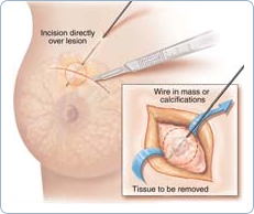
Surgical Biopsy
Sometimes, a needle is used to guide a surgical biopsy. A mammogram or ultrasound is used to find breast changes that cannot be felt. Once the breast change is found, a radiologist puts a needle into the breast so that the tip of the needle is at the area of breast change (called needle localization). Then a fine wire is placed through the needle. The needle may be removed and the wire stays in place. You may be asked to walk to another room or to the operating room for the biopsy. During surgery, the doctor uses the tip of the wire as a guide to what tissue to take out.
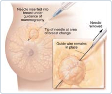
Needle Localization
Getting Your Biopsy Results
On average, you should get your biopsy results from your doctor 7 to 10 days after your biopsy.
The biopsy results will tell your doctor if you have:
- Benign breast changes
- Changes that may increase your chances of getting breast cancer
- Cancer cells in the lining of your breast ducts
- Cancer cells that have spread into nearby breast tissue
Here are more detailed descriptions of biopsy results.
Benign Changes
- Fibroadenomas--Hard, round lumps that feel like rubber
- Fat necrosis--Painless, round, firm lumps
- Sclerosing adenosis--Growths that may contain calcifications and are often painful
Your health care provider will probably suggest that you get follow-up care for these types of benign changes. See Chart 2: Possible Biopsy Results and Follow-Up Care for more information.
Two types of changes that may increase your chances of getting breast cancer
- Abnormal cells found in the breast ducts (Atypical Ductal Hyperplasia, or ADH)
- Abnormal cells found in the breast lobules (Lobular Carcinoma In Situ, or LCIS)
Jackie, Age 63
Atypical Ductal Hyperplasia (ADH) or Lobular Carcinoma In Situ (LCIS)
ADH and LCIS are not cancer. However, having ADH or LCIS in one breast does mean that you have a higher risk of getting cancer in either breast. Your doctor will want to follow your breast health very closely if you have ADH or LCIS. For example, your doctor may suggest that you:
- Get more mammograms or see him or her more often
- Take a drug, called tamoxifen, which has been shown to lower some women's risk of getting breast cancer
- Take part in clinical trials of promising new preventive treatments. In a cancer prevention trial, a large group of people is studied to help find better ways of preventing or lowering the chances of getting cancer.
A very small number of women with LCIS have surgery to remove both their breasts. They do this to try to keep breast cancer from developing. Talk to your doctor about what is best for you. See Chart 2: Possible Biopsy Results and Follow-Up Care for more information about ADH and LCIS.
Cancer Cells Found in the Lining of the Ducts (Ductal Carcinoma In Situ or DCIS)
DCIS is a condition in which cancer cells are only found inside the lining of a breast duct. The abnormal cells have not spread outside the duct to the surrounding breast tissue. Therefore, most women with DCIS are cured with treatment. However, if not treated, DCIS sometimes spreads to other parts of the breast (also called invasive breast cancer). Your doctor may discuss some of these treatment choices for DCIS:
- Getting breast-sparing surgery (the surgeon removes only your cancer and some normal tissue around it) with radiation therapy
- Getting breast-sparing surgery without radiation therapy
- Getting a total mastectomy (surgery to remove the entire breast)
- Taking the drug tamoxifen after surgery and radiation to lower the chances of the cancer coming back
See Chart 2: Possible Biopsy Results and Follow-Up Care for more information about DCIS.
Getting a Second Opinion
You can ask to have another pathologist review your biopsy results. This is important because biopsy results determine treatment(s) for breast changes and diseases. Ask your medical team to help you with this process.
Cancer That Is Invasive
Even if you have breast cancer that has spread to other parts of your breast, finding it early gives you the best chance to get treatment that may save your breasts and your life. It's also true that women with breast cancer that has spread have reasons for hope.
Some women with breast cancer may want to take part in a clinical trial. Clinical trials are research studies to find better ways to treat cancer. Some studies on new cancer-fighting drugs, drug combinations, and ways of giving treatment have already begun to show good results.
For more information about breast cancer and its treatments, call 1-800-4-CANCER for a free copy of What You Need To Know About Breast Cancer.
Finding the Support You Need
It can be scary when you find out about a breast change, get an abnormal mammogram result, or learn you have breast cancer. Many women have found it helpful to:
- Ask friends or loved ones for support. Take someone with you while you are learning about your testing and treatment choices.
- Ask your doctor or nurse to:
- Help you understand medical terms that are confusing
- Tell you how other people have handled the types of feelings that you are having
For More Information
The following organizations can give you more information on mammograms, breast changes, finding breast cancer early, and different breast cancer treatments.
National Cancer Institute (NCI)
You can find out more from these free NCI services:
Cancer Information Service
Toll free: 1-800-4-CANCER (1-800-422-6237)
TTY: 1-800-332-8615
- Answers to your questions about cancer
- Tips to prevent and screen for cancer
- Informational materials
- Other resources
This Web site provides current and accurate information from the National Cancer Institute. Through instant messaging (LiveHelp), Information Specialists can answer your questions about cancer and provide help in navigating NCI's Web site. You will find a wide range of cancer information, including:
- Treatment options
- Clinical trials
- Ways to reduce cancer risk
- Prevention and screening information
- Ways to cope with cancer
Centers for Medicare and Medicaid Services (CMS)
Toll free: 1-800-MEDICARE (1-800-633-4227)
TTY/TDD: 1-877-486-2048
Web site: www.cms.gov or www.medicare.gov
CMS provides information about Medicare benefits to pay for screening mammograms.
Centers for Disease Control and Prevention (CDC)
Toll free: 1-888-842-6355
Web site: www.cdc.gov/cancer
CDC conducts, supports, and promotes efforts to prevent cancer and increase early detection of cancer. CDC's National Breast and Cervical Cancer Early Detection Program provides free breast and cervical cancer screening and diagnostic services to women with low incomes.
National Women's Health Information Center (NWHIC)
Toll free: 1-800-994-WOMAN (1-800-994-9662)
TTY: 1-888-220-5446
Web site: www.4woman.gov
NWHIC provides a gateway to women's health information. NWHIC is sponsored by the U.S. Department of Health and Human Services Office on Women's Health.
U.S. Food and Drug Administration (FDA)
Web site: www.fda.gov/cdrh/mammography
The FDA has information about mammography on its Web site, including a section for consumers, a list of FDA-approved mammography facilities, and laws about how these facilities are run.
Chart 1: Possible Mammogram Results and Follow-Up Care
| Conditions | Features | What Your Doctor May Recommend |
Cysts |
|
|
Fibroadenoma |
|
|
Macrocalcifications |
|
|
Mass |
|
|
Microcalcifications |
|
|
Chart 2: Possible Biopsy Results and Follow-Up Care
| Conditions | Features | What Your Doctor May Recommend |
Atypical Ductal Hyperplasia (ADH) |
|
|
Cancer |
|
|
Ductal Carcinoma In Situ (DCIS) |
|
|
Fat Necrosis |
|
|
Fibroadenoma |
|
|
Intraductal Papilloma |
|
|
Lobular Carcinoma In Situ (LCIS) |
|
|
Sclerosing Adenosis |
|
|
More information on these conditions can be found on www.cancer.gov or by calling 1-800-4-CANCER.
Understanding Cancer
Cancer begins in cells, the building blocks that make up tissues. Tissues make up the organs of the body.
Normally, cells grow and divide to form new cells as the body needs them. When cells grow old, they die, and new cells take their place.
Sometimes, this orderly process goes wrong. New cells form when the body does not need them, and old cells do not die when they should. These extra cells can form a mass of tissue called a growth or tumor.
Tumors can be benign or malignant:
Benign tumors are not cancer:- Benign tumors are rarely life-threatening.
- Generally, benign tumors can be removed. They usually do not grow back.
- Cells from benign tumors do not invade the tissues around them.
- Cells from benign tumors do not spread to other parts of the body.
Malignant tumors are cancer:
- Malignant tumors are generally more serious than benign tumors. They may be life-threatening.
- Malignant tumors often can be removed. But sometimes they grow back.
- Cells from malignant tumors can invade and damage nearby tissues and organs.
- Cells from malignant tumors can spread (metastasize) to other parts of the body. Cancer cells spread by breaking away from the original (primary) tumor and entering the bloodstream or lymphatic system. The cells invade other organs and form new tumors that damage these organs. The spread of cancer is called metastasis.
When breast cancer cells spread, the cancer cells are often found in lymph nodes near the breast. Also, breast cancer can spread to almost any other part of the body. The most common are the bones, liver, lungs, and brain. The new tumor has the same kind of abnormal cells and the same name as the primary tumor. For example, if breast cancer spreads to the bones, the cancer cells in the bones are actually breast cancer cells. The disease is metastatic breast cancer, not bone cancer. For that reason, it is treated as breast cancer, not bone cancer. Doctors call the new tumor "distant" or metastatic disease.
Risk Factors
No one knows the exact causes of breast cancer. Doctors often cannot explain why one woman develops breast cancer and another does not. They do know that bumping, bruising, or touching the breast does not cause cancer. And breast cancer is not contagious. You cannot "catch" it from another person.
Research has shown that women with certain risk factors are more likely than others to develop breast cancer. A risk factor is something that may increase the chance of developing a disease.
Studies have found the following risk factors for breast cancer:
- Age: The chance of getting breast cancer goes up as a woman gets older. Most cases of breast cancer occur in women over 60. This disease is not common before menopause.
- Personal history of breast cancer: A woman who had breast cancer in one breast has an increased risk of getting cancer in her other breast.
- Family history: A woman's risk of breast cancer is higher if her mother, sister, or daughter had breast cancer. The risk is higher if her family member got breast cancer before age 40. Having other relatives with breast cancer (in either her mother's or father's family) may also increase a woman's risk.
- Certain breast changes: Some women have cells in the breast that look abnormal under a microscope. Having certain types of abnormal cells (atypical hyperplasia and lobular carcinoma in situ [LCIS]) increases the risk of breast cancer.
- Gene changes: Changes in certain genes increase the risk of breast cancer. These genes include BRCA1, BRCA2, and others. Tests can sometimes show the presence of specific gene changes in families with many women who have had breast cancer. Health care providers may suggest ways to try to reduce the risk of breast cancer, or to improve the detection of this disease in women who have these changes in their genes. NCI offers publications on gene testing.
Reproductive and menstrual history:
- The older a woman is when she has her first child, the greater her chance of breast cancer.
- Women who had their first menstrual period before age 12 are at an increased risk of breast cancer.
- Women who went through menopause after age 55 are at an increased risk of breast cancer.
- Women who never had children are at an increased risk of breast cancer.
- Women who take menopausal hormone therapy with estrogen plus progestin after menopause also appear to have » an increased risk of breast cancer.
- Large, well-designed studies have shown no link between abortion or miscarriage and breast cancer.
Race: Breast cancer is diagnosed more often in white women than Latina, Asian, or African American women.
Radiation therapy to the chest: Women who had radiation therapy to the chest (including breasts) before age 30 are at an increased risk of breast cancer. This includes women treated with radiation for Hodgkin's lymphoma. Studies show that the younger a woman was when she received radiation treatment, the higher her risk of breast cancer later in life.
Breast density: Breast tissue may be dense or fatty. Older women whose mammograms (breast x-rays) show more dense tissue are at increased risk of breast cancer.
Taking DES (diethylstilbestrol): DES was given to some pregnant women in the United States between about 1940 and 1971. (It is no longer given to pregnant women.) Women who took DES during pregnancy may have a slightly increased risk of breast cancer. The possible effects on their daughters are under study.
Being overweight or obese after menopause: The chance of getting breast cancer after menopause is higher in women who are overweight or obese.
Lack of physical activity: Women who are physically inactive throughout life may have an increased risk of breast cancer. Being active may help reduce risk by preventing weight gain and obesity.
Drinking alcohol: Studies suggest that the more alcohol a woman drinks, the greater her risk of breast cancer.
Other possible risk factors are under study. Researchers are studying the effect of diet, physical activity, and genetics on breast cancer risk. They are also studying whether certain substances in the environment can increase the risk of breast cancer.
Many risk factors can be avoided. Others, such as family history, cannot be avoided. Women can help protect themselves by staying away from known risk factors whenever possible.
But it is also important to keep in mind that most women who have known risk factors do not get breast cancer. Also, most women with breast cancer do not have a family history of the disease. In fact, except for growing older, most women with breast cancer have no clear risk factors.
If you think you may be at risk, you should discuss this concern with your doctor. Your doctor may be able to suggest ways to reduce your risk and can plan a schedule for checkups.
Screening
Screening Mammogram
Clinical Breast Exam
Breast Self-Exam
Screening for breast cancer before there are symptoms can be important. Screening can help doctors find and treat cancer early. Treatment is more likely to work well when cancer is found early.
Your doctor may suggest the following screening tests for breast cancer:
- Screening mammogram
- Clinical breast exam
- Breast self-exam
You should ask your doctor about when to start and how often to check for breast cancer.
Screening Mammogram
To find breast cancer early, NCI recommends that:
- Women in their 40s and older should have mammograms every 1 to 2 years. A mammogram is a picture of the breast made with x-rays.
- Women who are younger than 40 and have risk factors for breast cancer should ask their health care provider whether to have mammograms and how often to have them.
Mammograms can often show a breast lump before it can be felt. They also can show a cluster of tiny specks of calcium. These specks are called micro calcifications. Lumps or specks can be from cancer, precancerous cells, or other conditions. Further tests are needed to find out if abnormal cells are present.
If an abnormal area shows up on your mammogram, you may need to have more x-rays. You also may need a biopsy. A biopsy is the only way to tell for sure if cancer is present. (The "Diagnosis" section has more information on biopsy.)
Mammograms are the best tool doctors have to find breast cancer early. However, mammograms are not perfect:
- A mammogram may miss some cancers. (The result is called a "false negative.")
- A mammogram may show things that turn out not to be cancer. (The result is called a "false positive.")
- Some fast-growing tumors may grow large or spread to other parts of the body before a mammogram detects them.
Mammograms (as well as dental x-rays, and other routine x-rays) use very small doses of radiation. The risk of any harm is very slight, but repeated x-rays could cause problems. The benefits nearly always outweigh the risk. You should talk with your health care provider about the need for each x-ray. You should also ask for shields to protect parts of your body that are not in the picture.
Clinical Breast Exam
During a clinical breast exam, your health care provider checks your breasts. You may be asked to raise your arms over your head, let them hang by your sides, or press your hands against your hips.
Your health care provider looks for differences in size or shape between your breasts. The skin of your breasts is checked for a rash, dimpling, or other abnormal signs. Your nipples may be squeezed to check for fluid.
Using the pads of the fingers to feel for lumps, your health care provider checks your entire breast, underarm, and collarbone area. A lump is generally the size of a pea before anyone can feel it. The exam is done on one side, then the other. Your health care provider checks the lymph nodes near the breast to see if they are enlarged.
A thorough clinical breast exam may take about 10 minutes.
Breast Self-Exam
You may perform monthly breast self-exams to check for any changes in your breasts. It is important to remember that changes can occur because of aging, your menstrual cycle, pregnancy, menopause, or taking birth control pills or other hormones. It is normal for breasts to feel a little lumpy and uneven. Also, it is common for your breasts to be swollen and tender right before or during your menstrual period.
You should contact your health care provider if you notice any unusual changes in your breasts.
Breast self-exams cannot replace regular screening mammograms and clinical breast exams. Studies have not shown that breast self-exams alone reduce the number of deaths from breast cancer.
You may want to ask the doctor the following questions about screening:
- Which tests do you recommend for me? Why?
- Do the tests hurt? Are there any risks?
- How much do mammograms cost? Will my health insurance pay for them?
- How soon after the mammogram will I learn the results?
- If the results show a problem, how will you learn if I have cancer?
Symptoms
Common symptoms of breast cancer include:
» A change in how the breast or nipple feels
- A lump or thickening in or near the breast or in the underarm area
- Nipple tenderness
» A change in how the breast or nipple looks
- A change in the size or shape of the breast
- A nipple turned inward into the breast
- The skin of the breast, areola, or nipple may be scaly, red, or swollen. It may have ridges or pitting so that it looks like the skin of an orange.
» Nipple discharge (fluid)
Early breast cancer usually does not cause pain. Still, a woman should see her health care provider about breast pain or any other symptom that does not go away. Most often, these symptoms are not due to cancer. Other health problems may also cause them. Any woman with these symptoms should tell her doctor so that problems can be diagnosed and treated as early as possible
Diagnosis
If you have a symptom or screening test result that suggests cancer, your doctor must find out whether it is due to cancer or to some other cause. Your doctor may ask about your personal and family medical history. You may have a physical exam. Your doctor also may order a mammogram or other imaging procedure. These tests make pictures of tissues inside the breast. After the tests, your doctor may decide no other exams are needed. Your doctor may suggest that you have a follow-up exam later on. Or you may need to have a biopsy to look for cancer cells.
Clinical Breast Exam
Your health care provider feels each breast for lumps and looks for other problems. If you have a lump, your doctor will feel its size, shape, and texture. Your doctor will also check to see if it moves easily. Benign lumps often feel different from cancerous ones. Lumps that are soft, smooth, round, and movable are likely to be benign. A hard, oddly shaped lump that feels firmly attached within the breast is more likely to be cancer.
Diagnostic Mammogram
Diagnostic mammograms are x-ray pictures of the breast. They take clearer, more detailed images of areas that look abnormal on a screening mammogram. Doctors use them to learn more about unusual breast changes, such as a lump, pain, thickening, nipple discharge, or change in breast size or shape. Diagnostic mammograms may focus on a specific area of the breast. They may involve special techniques and more views than screening mammograms.
Ultrasound
An ultrasound device sends out sound waves that people cannot hear. The waves bounce off tissues. A computer uses the echoes to create a picture. Your doctor can view these pictures on a monitor. The pictures may show whether a lump is solid or filled with fluid. A cyst is a fluid-filled sac. Cysts are not cancer. But a solid mass may be cancer. After the test, your doctor can store the pictures on video or print them out. This exam may be used along with a mammogram.
Magnetic Resonance Imaging
Magnetic resonance imaging (MRI) uses a powerful magnet linked to a computer. MRI makes detailed pictures of breast tissue. Your doctor can view these pictures on a monitor or print them on film. MRI may be used along with a mammogram.
Biopsy
Your doctor may refer you to a surgeon or breast disease specialist for a biopsy. Fluid or tissue is removed from your breast to help find out if there is cancer.
Some suspicious areas can be seen on a mammogram but cannot be felt during a clinical breast exam. Doctors can use imaging procedures to help see the area and remove tissue. Such procedures include ultrasound-guided, needle-localized, or stereotactic biopsy.
Doctors can remove tissue from the breast in different ways:
Fine-needle aspiration: Your doctor uses a thin needle to remove fluid from a breast lump. If the fluid appears to contain cells, a pathologist at a lab checks them for cancer with a microscope. If the fluid is clear, it may not need to be checked by a lab.
Core biopsy: Your doctor uses a thick needle to remove breast tissue. A pathologist checks for cancer cells. This procedure is also called a needle biopsy.
Surgical biopsy: Your surgeon removes a sample of tissue. A pathologist checks the tissue for cancer cells.
- An incisional biopsy takes a sample of a lump or abnormal area.
- An excisional biopsy takes the entire lump or area.
If cancer cells are found, the pathologist can tell what kind of cancer it is. The most common type of breast cancer is ductal carcinoma. Abnormal cells are found in the lining of the ducts. Lobular carcinoma is another type. Abnormal cells are found in the lobules.
You may want to ask your doctor the following questions before having a biopsy:
- What kind of biopsy will I have? Why?
- How long will it take? Will I be awake? Will it hurt? Will I have anesthesia? What kind?
- Are there any risks? What are the chances of infection or bleeding after the biopsy?
- How soon will I know the results?
- If I do have cancer, who will talk with me about the next steps? When?
Additional Tests
If you are diagnosed with cancer, your doctor may order special lab tests on the breast tissue that was removed. These tests help your doctor learn more about the cancer and plan treatment:
Hormone receptor test: This test shows whether the tissue has certain hormone receptors. Tissue with these receptors needs hormones (estrogen or progesterone) to grow.
HER2 test: This test shows whether the tissue has a protein called human epidermal growth factor receptor-2 (HER2) or the HER2/neu gene. Having too much protein or too many copies of the gene in the tissue may increase the chance that the breast cancer will come back after treatment.
Staging
To plan your treatment, your doctor needs to know the extent (stage) of the disease. The stage is based on the size of the tumor and whether the cancer has spread. Staging may involve x-rays and lab tests. These tests can show whether the cancer has spread and, if so, to what parts of your body. When breast cancer spreads, cancer cells are often found in lymph nodes under the arm (axillary lymph nodes). The stage often is not known until after surgery to remove the tumor in your breast and the lymph nodes under your arm.
These are the stages of breast cancer:
- Stage 0 is carcinoma in situ
- Lobular carcinoma in situ (LCIS): Abnormal cells are in the lining of a lobule. (See picture of lobule on page 3.) LCIS seldom becomes invasive cancer. However, having LCIS in one breast increases the risk of cancer for both breasts.
- Ductal carcinoma in situ (DCIS): Abnormal cells are in the lining of a duct. DCIS is also called intraductal carcinoma .The abnormal cells have not spread outside the duct. They have not invaded the nearby breast tissue. DCIS sometimes becomes invasive cancer if not treated.
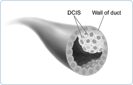
This picture shows ductal carcinoma in situ. - Stage I is an early stage of invasive breast cancer. The tumor is no more than 2 centimeters (three-quarters of an inch) across. Cancer cells have not spread beyond the breast.
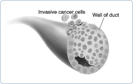
This picture shows cancer cells spreading outside the duct. The cancer cells are invading nearby tissue inside the breast. - Stage II is one of the following:
- The tumor is no more than 2 centimeters (three-quarters of an inch) across. The cancer has spread to the lymph nodes under the arm.
- The tumor is between 2 and 5 centimeters (three-quarters of an inch to 2 inches). The cancer has not spread to the lymph nodes under the arm.
- The tumor is between 2 and 5 centimeters (three-quarters of an inch to 2 inches). The cancer has spread to the lymph nodes under the arm
- The tumor is larger than 5 centimeters (2 inches). The cancer has not spread to the lymph nodes under the arm.
- Stage III is locally advanced cancer. It is divided into Stage IIIA, IIIB, and IIIC
- Stage IIIA is one of the following:
- The tumor is no more than 5 centimeters (2 inches) across. The cancer has spread to underarm lymph nodes that are attached to each other or to other structures. Or the cancer may have spread to lymph nodes behind the breastbone.
- The tumor is more than 5 centimeters across. The cancer has spread to underarm lymph nodes that are either alone or attached to each other or to other structures. Or the cancer may have spread to lymph nodes behind the breastbone.
- Stage IIIB is a tumor of any size that has grown into the chest wall or the skin of the breast. It may be associated with swelling of the breast or with nodules (lumps) in the breast skin.
- The cancer may have spread to lymph nodes under the arm.
- The cancer may have spread to underarm lymph nodes that are attached to each other or other structures. Or the cancer may have spread to lymph nodes behind the breastbone.
- Inflammatory breast cancer is a rare type of breast cancer. The breast looks red and swollen because cancer cells block the lymph vessels in the skin of the breast. When a doctor diagnoses inflammatory breast cancer, it is at least Stage IIIB, but it could be more advanced.
- Stage IIIC is a tumor of any size. It has spread in one of the following ways:
- The cancer has spread to the lymph nodes behind the breastbone and under the arm.
- The cancer has spread to the lymph nodes above or below the collarbone.
- Stage IV is distant metastatic cancer. The cancer has spread to other parts of the body.
Recurrent cancer is cancer that has come back (recurred) after a period of time when it could not be detected. It may recur locally in the breast or chest wall. Or it may recur in any other part of the body, such as the bone, liver, or lungs.
Treatment
» Getting a Second Opinion
» Treatment Methods
Many women with breast cancer want to take an active part in making decisions about their medical care. It is natural to want to learn all you can about your disease and treatment choices. Knowing more about breast cancer helps many women cope.
Shock and stress after the diagnosis can make it hard to think of everything you want to ask your doctor. It often helps to make a list of questions before an appointment. To help remember what the doctor says, you may take notes or ask whether you may use a tape recorder. You may also want to have a family member or friend with you when you talk to the doctor - to take part in the discussion, to take notes, or just to listen. You do not need to ask all your questions at once. You will have other chances to ask your doctor or nurse to explain things that are not clear and to ask for more details.
Your doctor may refer you to a specialist, or you may ask for a referral. Specialists who treat breast cancer include surgeons, medical oncologists, and radiation oncologists. You also may be referred to a plastic surgeon.
Getting a Second Opinion
Before starting treatment, you might want a second opinion about your diagnosis and treatment plan. Many insurance companies cover a second opinion if you or your doctor requests it. It may take some time and effort to gather medical records and arrange to see another doctor. You may have to gather your mammogram films, biopsy slides, pathology report, and proposed treatment plan. Usually it is not a problem to take several weeks to get a second opinion. In most cases, the delay in starting treatment will not make treatment less effective. To make sure, you should discuss this delay with your doctor. Some women with breast cancer need treatment right away.
There are a number of ways to find a doctor for a second opinion:
- Your doctor may refer you to one or more specialists. At cancer centers, several specialists often work together as a team.
- A local or state medical society, a nearby hospital, or a medical school can usually provide the names of specialists.
- The American Board of Medical Specialties (ABMS) has a list of doctors who have had training and passed exams in their specialty. You can find this list in the Official ABMS Directory of Board Certified Medical Specialists. This Directory is in most public libraries. Also, ABMS offers this information at http://www.abms.org. (Click on "Who's Certified.")
- NCI provides a helpful fact sheet called "How to Find a Doctor or Treatment Facility If You Have Cancer."
Treatment Methods
Women with breast cancer have many treatment options. These include surgery, radiation therapy, chemotherapy, hormone therapy, and biological therapy. These options are described below. Many women receive more than one type of treatment.
The choice of treatment depends mainly on the stage of the disease. Treatment options by stage are described below.
Your doctor can describe your treatment choices and the expected results. You may want to know how treatment may change your normal activities. You may want to know how you will look during and after treatment. You and your doctor can work together to develop a treatment plan that reflects your medical needs and personal values.
Cancer treatment is either local therapy or systemic therapy:
- Local therapy: Surgery and radiation therapy are local treatments. They remove or destroy cancer in the breast. When breast cancer has spread to other parts of the body, local therapy may be used to control the disease in those specific areas.
- Systemic therapy: Chemotherapy, hormone therapy, and biological therapy are systemic treatments. They enter the bloodstream and destroy or control cancer throughout the body. Some women with breast cancer have systemic therapy to shrink the tumor before surgery or radiation. Others have systemic therapy after surgery and/or radiation to prevent the cancer from coming back. Systemic treatments also are used for cancer that has spread.
Because cancer treatments often damage healthy cells and tissues, side effects are common. Side effects depend mainly on the type and extent of the treatment. Side effects may not be the same for each woman, and they may change from one treatment session to the next.
Before treatment starts, your health care team will explain possible side effects and suggest ways to help you manage them. There are many helpful booklets about cancer treatments and coping with side effects. These include Radiation Therapy and You, Chemotherapy and You, Biological Therapy, and Eating Hints for Cancer Patients.
At any stage of disease, supportive care is available to control pain and other symptoms, to relieve the side effects of treatment, and to ease emotional concerns.
You may want to ask your doctor these questions before your treatment begins:
- What did the hormone receptor test show? What did other lab tests show?
- Do any lymph nodes show signs of cancer?
- What is the stage of the disease? Has the cancer spread?
- What is the goal of treatment? What are my treatment choices? Which do you recommend for me? Why?
- What are the expected benefits of each kind of treatment?
- What are the risks and possible side effects of each treatment? How can side effects be managed?
- What can I do to prepare for treatment?
- Will I need to stay in the hospital? If so, for how long?
- What is the treatment likely to cost? Will my insurance cover the cost?
- How will treatment affect my normal activities?
- Would a clinical trial be appropriate for me?
Surgery
Surgery is the most common treatment for breast cancer. There are several types of surgery. (See pictures below.) Your doctor can explain each type, discuss and compare the benefits and risks, and describe how each will change the way you look:
- Breast-sparing surgery: An operation to remove the cancer but not the breast is breast-sparing surgery. It is also called breast-conserving surgery, lumpectomy, segmental mastectomy, and partial mastectomy. Sometimes an excisional biopsy serves as a lumpectomy because the surgeon removes the whole lump.
The surgeon often removes the underarm lymph nodes as well. A separate incision is made. This procedure is called an axillary lymph node dissection. It shows whether cancer cells have entered the lymphatic system.
After breast-sparing surgery, most women receive radiation therapy to the breast. This treatment destroys cancer cells that may remain in the breast. - Mastectomy: An operation to remove the breast (or as much of the breast tissue as possible) is a mastectomy. In most cases, the surgeon also removes lymph nodes under the arm. Some women have radiation therapy after surgery.
Studies have found equal survival rates for breast-sparing surgery (with radiation therapy) and mastectomy for Stage I and Stage II breast cancer.
Sentinel lymph node biopsy is a new method of checking for cancer cells in the lymph nodes. A surgeon removes fewer lymph nodes, which causes fewer side effects. (If the doctor finds cancer cells in the axillary lymph nodes, an axillary lymph node dissection usually is done.) Information about ongoing studies of sentinel lymph node biopsy is in the section on "The Promise of Cancer Research." These studies will learn the lasting effects of removing fewer lymph nodes.
In breast-sparing surgery, the surgeon removes the tumor in the breast and some tissue around it. The surgeon may also remove lymph nodes under the arm. The surgeon sometimes removes some of the lining over the chest muscles below the tumor.

In total (simple) mastectomy, the surgeon removes the whole breast. Some lymph nodes under the arm may also be removed.

In modified radical mastectomy, the surgeon removes the whole breast, and most or all of the lymph nodes under the arm. Often, the lining over the chest muscles is removed. A small chest muscle also may be taken out to make it easier to remove the lymph nodes.
You may choose to have breast reconstruction. This is plastic surgery to rebuild the shape of the breast. It may be done at the same time as a mastectomy or later. If you are considering reconstruction, you may wish to talk with a plastic surgeon before having a mastectomy. More information is in the "Breast Reconstruction" section.
The time it takes to heal after surgery is different for each woman. Surgery causes pain and tenderness. Medicine can help control the pain. Before surgery, you should discuss the plan for pain relief with your doctor or nurse. After surgery, your doctor can adjust the plan if you need more relief. Any kind of surgery also carries a risk of infection, bleeding, or other problems. You should tell your health care provider right away if you develop any problems.
The time it takes to heal after surgery is different for each woman. Surgery causes pain and tenderness. Medicine can help control the pain. Before surgery, you should discuss the plan for pain relief with your doctor or nurse. After surgery, your doctor can adjust the plan if you need more relief. Any kind of surgery also carries a risk of infection, bleeding, or other problems. You should tell your health care provider right away if you develop any problems.
Because nerves may be injured or cut during surgery, you may have numbness and tingling in your chest, underarm, shoulder, and upper arm. These feelings usually go away within a few weeks or months. But for some women, numbness does not go away.
Removing the lymph nodes under the arm slows the flow of lymph fluid. The fluid may build up in your arm and hand and cause swelling. This swelling is lymphedema. Lymphedema can develop right after surgery or months to years later.
You will need to protect your arm and hand on the treated side for the rest of your life:
- Avoid wearing tight clothing or jewelry on your affected arm
- Carry your purse or luggage with the other arm
- Use an electric razor to avoid cuts when shaving under your arm
- Have shots, blood tests, and blood pressure measurements on the other arm
- Wear gloves to protect your hands when gardening and when using strong detergents
- Have careful manicures and avoid cutting your cuticles
- Avoid burns or sunburns to your affected arm and hand
You should ask your doctor how to handle any cuts, insect bites, sunburn, or other injuries to your arm or hand. Also, you should contact the doctor if your arm or hand is injured, swells, or becomes red and warm.
If lymphedema occurs, the doctor may suggest raising your arm above your heart whenever you can. The doctor may show you hand and arm exercises. Some women with lymphedema wear an elastic sleeve to improve lymph circulation. Medication, manual lymph drainage (massage), or use of a machine that gently compresses the arm may also help. You may be referred to a physical therapist or another specialist.
You may want to ask your doctor these questions before having surgery:
- What kinds of surgery can I consider? Is breast-sparing surgery an option for me? Which operation do you recommend for me? Why?
- Will my lymph nodes be removed? How many? Why?
- How will I feel after the operation? Will I have to stay in the hospital?
- Will I need to learn how to take care of myself or my incision when I get home?
- Where will the scars be? What will they look like?
- If I decide to have plastic surgery to rebuild my breast, how and when can that be done? Can you suggest a plastic surgeon for me to contact?
- Will I have to do special exercises to help regain motion and strength in my arm and shoulder? Will a physical therapist or nurse show me how to do the exercises?
- Is there someone I can talk with who has had the same surgery I'll be having?
Radiation Therapy
Radiation therapy (also called radiotherapy) uses high-energy rays to kill cancer cells. Most women receive radiation therapy after breast-sparing surgery. Some women receive radiation therapy after a mastectomy. Treatment depends on the size of the tumor and other factors. The radiation destroys breast cancer cells that may remain in the area.
Some women have radiation therapy before surgery to destroy cancer cells and shrink the tumor. Doctors use this approach when the tumor is large or may be hard to remove. Some women also have chemotherapy or hormone therapy before surgery.
Doctors use two types of radiation therapy to treat breast cancer. Some women receive both types:
- External radiation: The radiation comes from a large machine outside the body. Most women go to a hospital or clinic for treatment. Treatments are usually 5 days a week for several weeks.
- Internal radiation (implant radiation): Thin plastic tubes (implants) that hold a radioactive substance are put directly in the breast. The implants stay in place for several days. A woman stays in the hospital while she has implants. Doctors remove the implants before she goes home.
Side effects depend mainly on the dose and type of radiation and the part of your body that is treated.
It is common for the skin in the treated area to become red, dry, tender, and itchy. Your breast may feel heavy and tight. These problems will go away over time. Toward the end of treatment, your skin may become moist and "weepy." Exposing this area to air as much as possible can help the skin heal.
Bras and some other types of clothing may rub your skin and cause soreness. You may want to wear loose-fitting cotton clothes during this time. Gentle skin care also is important. You should check with your doctor before using any deodorants, lotions, or creams on the treated area. These effects of radiation therapy on the skin will go away. The area gradually heals once treatment is over. However, there may be a lasting change in the color of your skin.
You are likely to become very tired during radiation therapy, especially in the later weeks of treatment. Resting is important, but doctors usually advise patients to try to stay as active as they can.
Although the side effects of radiation therapy can be distressing, your doctor can usually relieve them.
You may want to ask your doctor these questions before having radiation therapy:
- How will radiation be given?
- When will treatment start? When will it end? How often will I have treatments?
- How will I feel during treatment? Will I be able to drive myself to and from treatment?
- How will we know the treatment is working?
- What can I do to take care of myself before, during, and after treatment?
- Will treatment affect my skin?
- How will my chest look afterward?
- Are there any long-term effects?
- What is the chance that the cancer will come back in my breast?
- How often will I need checkups?
Chemotherapy
Chemotherapy uses anticancer drugs to kill cancer cells. Chemotherapy for breast cancer is usually a combination of drugs. The drugs may be given as a pill or by injection into a vein (IV). Either way, the drugs enter the bloodstream and travel throughout the body.
Women with breast cancer can have chemotherapy in an outpatient part of the hospital, at the doctor's office, or at home. Some women need to stay in the hospital during treatment.
Side effects depend mainly on the specific drugs and the dose. The drugs affect cancer cells and other cells that divide rapidly:
- Blood cells: These cells fight infection, help your blood to clot, and carry oxygen to all parts of the body. When drugs affect your blood cells, you are more likely to get infections, bruise or bleed easily, and feel very weak and tired. Years after chemotherapy, some women have developed leukemia (cancer of the blood cells).
- Cells in hair roots: Chemotherapy can cause hair loss. Your hair will grow back, but it may be somewhat different in color and texture.
- Cells that line the digestive tract: Chemotherapy can cause poor appetite, nausea and vomiting, diarrhea, or mouth and lip sores.
Your doctor can suggest ways to control many of these side effects.
Some drugs used for breast cancer can cause tingling or numbness in the hands or feet. This problem usually goes away after treatment is over. Other problems may not go away. In some women, the drugs used for breast cancer may weaken the heart.
Some anticancer drugs can damage the ovaries. The ovaries may stop making hormones. You may have symptoms of menopause. The symptoms include hot flashes and vaginal dryness. Your menstrual periods may no longer be regular or may stop. Some women become infertile (unable to become pregnant). For women over the age of 35, infertility is likely to be permanent.
On the other hand, you may remain fertile during chemotherapy and be able to become pregnant. The effects of chemotherapy on an unborn child are not known. You should talk to your doctor about birth control before treatment begins.
Hormone Therapy
Some breast tumors need hormones to grow. Hormone therapy keeps cancer cells from getting or using the natural hormones they need. These hormones are estrogen and progesterone. Lab tests can show if a breast tumor has hormone receptors. If you have this kind of tumor, you may have hormone therapy.
This treatment uses drugs or surgery:
- Drugs: Your doctor may suggest a drug that can block the natural hormone. One drug is tamoxifen, which blocks estrogen. Another type of drug prevents the body from making the female hormone estradiol. Estradiol is a form of estrogen. This type of drug is an aromatase inhibitor. If you have not gone through menopause, your doctor may give you a drug that stops the ovaries from making estrogen.
- Surgery: If you have not gone through menopause, you may have surgery to remove your ovaries. The ovaries are the main source of the body's estrogen. A woman who has gone through menopause does not need surgery. (The ovaries produce less estrogen after menopause.)
The side effects of hormone therapy depend largely on the specific drug or type of treatment. Tamoxifen is the most common hormone treatment. In general, the side effects of tamoxifen are similar to some of the symptoms of menopause. The most common are hot flashes and vaginal discharge. Other side effects are irregular menstrual periods, headaches, fatigue, nausea, vomiting, vaginal dryness or itching, irritation of the skin around the vagina, and skin rash. Not all women who take tamoxifen have side effects.
It is possible to become pregnant when taking tamoxifen. Tamoxifen may harm the unborn baby. If you are still menstruating, you should discuss birth control methods with your doctor.
Serious side effects of tamoxifen are rare. However, it can cause blood clots in the veins. Blood clots form most often in the legs and in the lungs. Women have a slight increase in their risk of stroke.
Tamoxifen can cause cancer of the uterus. Your doctor should perform regular pelvic exams. You should tell your doctor about any unusual vaginal bleeding between exams.
When the ovaries are removed, menopause occurs at once. The side effects are often more severe than those caused by natural menopause. Your health care provider can suggest ways to cope with these side effects.
Biological Therapy
Biological therapy helps the immune system fight cancer. The immune system is the body's natural defense against disease.
Some women with breast cancer that has spread receive a biological therapy called Herceptin® (trastuzumab). It is a monoclonal antibody. It is made in the laboratory and binds to cancer cells.
Herceptin is given to women whose lab tests show that a breast tumor has too much of a specific protein known as HER2. By blocking HER2, it can slow or stop the growth of the cancer cells.
Herceptin is given by vein. It may be given alone or with chemotherapy.
The first time a woman receives Herceptin, the most common side effects are fever and chills. Some women also have pain, weakness, nausea, vomiting, diarrhea, headaches, difficulty breathing, or rashes. Side effects usually become milder after the first treatment.
Herceptin also may cause heart damage. This may lead to heart failure. Herceptin can also affect the lungs. It can cause breathing problems that require a doctor at once. Before you receive Herceptin, your doctor will check for your heart and lungs. During treatment, your doctor will watch for signs of lung problems.
You may want to ask your doctor these questions before having chemotherapy, hormone therapy, or biological therapy:
- What drugs will I be taking? What will they do?
- If I need hormone treatment, would you recommend drugs or surgery to remove my ovaries?
- When will treatment start? When will it end? How often will I have treatments?
- Where will I go for treatment? Will I be able to drive home afterward?
- What can I do to take care of myself during treatment?
- How will we know the treatment is working?
- Which side effects should I tell you about?
- Will there be long-term effects?
Treatment Choices by Stage
Your treatment options depend on the stage of your disease and these factors:
- The size of the tumor in relation to the size of your breast
- The results of lab tests (such as whether the breast cancer cells need hormones to grow)
- Whether you have gone through menopause
- Your general health
Below are brief descriptions of common treatments for each stage. Other treatments may be appropriate for some women.
Stage 0
Stage 0 breast cancer refers to lobular carcinoma in situ (LCIS) or ductal carcinoma in situ (DCIS):
- LCIS: Most women with LCIS do not have treatment. Instead, the doctor may suggest regular checkups to watch for signs of breast cancer.
Some women take tamoxifen to reduce the risk of developing breast cancer. Others may take part in studies of promising new preventive treatments.
Having LCIS in one breast increases the risk of cancer for both breasts. A very small number of women with LCIS try to prevent cancer with surgery to remove both breasts. This is a bilateral prophylactic mastectomy. The surgeon usually does not remove the underarm lymph nodes.
- DCIS: Most women with DCIS have breast-sparing surgery followed by radiation therapy. Some women choose to have a total mastectomy. Underarm lymph nodes are not usually removed. Women with DCIS may receive tamoxifen to reduce the risk of developing invasive breast cancer.
Stages I, II, IIIA, and Operable IIIC
Women with Stage I, II, IIIA, and operable (can treat with surgery) IIIC breast cancer may have a combination of treatments. Some may have breast-sparing surgery followed by radiation therapy to the breast. This choice is common for women with Stage I or II breast cancer. Others decide to have a mastectomy.
With either approach, women (especially those with Stage II or IIIA breast cancer) often have lymph nodes under the arm removed. The doctor may suggest radiation therapy after mastectomy if cancer cells are found in 1 to 3 lymph nodes under the arm, or if the tumor in the breast is large. If cancer cells are found in more than 3 lymph nodes under the arm, the doctor usually will suggest radiation therapy after mastectomy.
The choice between breast-sparing surgery (followed by radiation therapy) and mastectomy depends on many factors:
- The size, location, and stage of the tumor
- The size of the woman's breast
- Certain features of the cancer
- How the woman feels about saving her breast
- How the woman feels about radiation therapy
- The woman's ability to travel to a radiation treatment center
Some women have chemotherapy before surgery. This is neoadjuvant therapy (treatment before the main treatment). Chemotherapy before surgery may shrink a large tumor so that breast-sparing surgery is possible. Women with large Stage II or IIIA breast tumors often choose this treatment.
After surgery, many women receive adjuvant therapy. Adjuvant therapy is treatment given after the main treatment to increase the chances of a cure. Radiation treatment can kill cancer cells in and near the breast. Women also may have systemic treatment such as chemotherapy, hormone therapy, or both. This treatment can destroy cancer cells that remain anywhere in the body. It can prevent the cancer from coming back in the breast or elsewhere.
Stages IIIB and Inoperable IIIC
Women with Stage IIIB (including inflammatory breast cancer) or inoperable Stage IIIC breast cancer usually have chemotherapy. (Inoperable cancer means it cannot be treated with surgery.)
If the chemotherapy shrinks the tumor, the doctor then may suggest further treatment:
- Mastectomy: The surgeon removes the breast. In most cases, the lymph nodes under the arm are removed. After surgery, a woman may receive radiation therapy to the chest and underarm area.
- Breast-sparing surgery: The surgeon removes the cancer but not the breast. In most cases, the lymph nodes under the arm are removed. After surgery, a woman may receive radiation therapy to the breast and underarm area.
- Radiation therapy instead of surgery: Some women have radiation therapy but no surgery. The doctor also may recommend more chemotherapy, hormone therapy, or both. This therapy may help prevent the disease from coming back in the breast or elsewhere.
Stage IV
In most cases, women with Stage IV breast cancer have hormone therapy, chemotherapy, or both. Some also may have biological therapy. Radiation may be used to control tumors in certain parts of the body. These treatments are not likely to cure the disease, but they may help a woman live longer.
Many women have supportive care along with anticancer treatments. Anticancer treatments are given to slow the progress of the disease. Supportive care helps manage pain, other symptoms, or side effects (such as nausea). It does not aim to extend a woman's life. Supportive care can help a woman feel better physically and emotionally. Some women with advanced cancer decide to have only supportive care.
Recurrent Breast Cancer
Recurrent cancer is cancer that has come back after it could not be detected. Treatment for the recurrent disease depends mainly on the location and extent of the cancer. Another main factor is the type of treatment the woman had before.
If breast cancer comes back only in the breast after breast-sparing surgery, the woman may have a mastectomy. Chances are good that the disease will not come back again.
If breast cancer recurs in other parts of the body, treatment may involve chemotherapy, hormone therapy, or biological therapy. Radiation therapy may help control cancer that recurs in the chest muscles or in certain other areas of the body.
Treatment can seldom cure cancer that recurs outside the breast. Supportive care is often an important part of the treatment plan. Many patients have supportive care to ease their symptoms and anticancer treatments to slow the progress of the disease. Some receive only supportive care to improve their quality of life.
Breast Reconstruction
Some women who plan to have a mastectomy decide to have breast reconstruction. Other women prefer to wear a breast form (prosthesis). Others decide to do nothing. All of these options have pros and cons. What is right for one woman may not be right for another. What is important is that nearly every woman treated for breast cancer has choices.
Breast reconstruction may be done at the same time as the mastectomy, or later on. If you are thinking about breast reconstruction, you should talk to a plastic surgeon before the mastectomy, even if you plan to have your reconstruction later on.
There are many ways to reconstruct the breast. Some women choose to have implants. Implants may be made of saline or silicone. The safety of silicone breast implants has been under review by the Food and Drug Administration (FDA) for several years. If you are thinking about having silicone implants, you may want to talk with your doctor about the FDA findings. Your doctor can tell you if silicone implants are an option.
You also may have breast reconstruction with tissue that the plastic surgeon moves from another part of your body. Skin, muscle, and fat can come from your lower abdomen, back, or buttocks. The surgeon uses this tissue to create a breast shape.
Which type of reconstruction is best depends on your age, body type, and the type of surgery you had. The plastic surgeon can explain the risks and benefits of each type of reconstruction.
You may want to ask your doctor these questions about breast reconstruction:
- What is the latest information about the safety of silicone breast implants?
- Which type of surgery would give me the best results? How will I look afterward?
- When can my reconstruction begin?
- How many surgeries will I need?
- What are the risks at the time of surgery? Later?
- Will I have scars? Where? What will they look like?
- If tissue from another part of my body is used, will there be any permanent changes where the tissue was removed?
- What activities should I avoid? When can I return to my normal activities?
- Will I need follow-up care?
- How much will reconstruction cost? Will my health insurance pay for it?
Complementary and Alternative Medicine
Some women with breast cancer use complementary and alternative medicine (CAM): Forms of treatment that are used in addition to (complementary) or instead of (alternative) standard treatments. These practices generally are not considered standard medical approaches. Standard treatments go through a long and careful research process to prove they are safe and effective, but less is known about most types of CAM. CAM may include dietary supplements, megadose vitamins, herbal preparations, special teas, acupuncture, massage therapy, magnet therapy, spiritual healing, and meditation.
- An approach is generally called complementary medicine when it is used along with standard treatment.
- An approach is called alternative medicine when it is used instead of standard treatment.
You may want to ask the doctor these questions before you decide to use CAM:
- What benefits can I expect from this approach?
- What are its risks?
- Do the expected benefits outweigh the risks?
- What side effects should I watch for?
- Will the approach change the way my cancer treatment works? Could this be harmful?
- Is this approach under study in a clinical trial? If so, who sponsors the trial?
- Will my health insurance pay for this approach?
Acupuncture, massage therapy, herbal products, vitamins or special diets, visualization, meditation, and spiritual healing are types of CAM.
Many women say that CAM helps them feel better. However, some types of CAM may change the way standard treatment works. These changes could be harmful. And some types of CAM could be harmful even if used alone.
Some types of CAM are expensive. Health insurance may not cover the cost.
Nutrition and Physical Activity
It is important for women with breast cancer to take care of themselves. Taking care of yourself includes eating well and staying as active as you can.
You need the right amount of calories to maintain a good weight. You also need enough protein to keep up your strength. Eating well may help you feel better and have more energy.
Sometimes, especially during or soon after treatment, you may not feel like eating. You may be uncomfortable or tired. You may find that foods do not taste as good as they used to. In addition, the side effects of treatment (such as poor appetite, nausea, vomiting, or mouth sores) can make it hard to eat well. Your doctor, dietitian, or other health care provider can suggest ways to deal with these problems.
Many women find they feel better when they stay active. Walking, yoga, swimming, and other activities can keep you strong and increase your energy. Exercise may reduce nausea and pain and make treatment easier to handle. It also can help relieve stress. Whatever physical activity you choose, be sure to talk to your doctor before you start. Also, if your activity causes you pain or other problems, be sure to let your doctor or nurse know about it.
Follow-up Care
Follow-up care after treatment for breast cancer is important. Recovery is different for each woman. Your recovery depends on your treatment, whether the disease has spread, and other factors.
Even when the cancer seems to have been completely removed or destroyed, the disease sometimes returns because undetected cancer cells remained somewhere in the body after treatment. Your doctor will monitor your recovery and check for recurrence of the cancer.
You should report any changes in the treated area or in your other breast to the doctor right away. Tell your doctor about any health problems, such as pain, loss of appetite or weight, changes in menstrual cycles, unusual vaginal bleeding, or blurred vision. Also talk to your doctor about headaches, dizziness, shortness of breath, coughing or hoarseness, backaches, or digestive problems that seem unusual or that don't go away. Such problems may arise months or years after treatment. They may suggest that the cancer has returned, but they can also be symptoms of other health problems. It is important to share your concerns with your doctor so problems can be diagnosed and treated as soon as possible.
Follow-up exams usually include the breasts, chest, neck, and underarm areas. Since you are at risk of getting cancer again, you should have mammograms of your preserved breast and your other breast. You probably will not need a mammogram of a reconstructed breast or if you had a mastectomy without reconstruction. Your doctor may order other imaging procedures or lab tests.
Sources of Support
Learning you have breast cancer can change your life and the lives of those close to you. These changes can be hard to handle. It is normal for you, your family, and your friends to have many different and sometimes confusing feelings.
You may worry about caring for your family, keeping your job, or continuing daily activities. Concerns about treatments and managing side effects, hospital stays, and medical bills are also common. Doctors, nurses, and other members of the health care team can answer questions about treatment, working, or other activities. Meeting with a social worker, counselor, or member of the clergy can be helpful if you want to talk about your feelings or concerns. Often, a social worker can suggest resources for financial aid, transportation, home care, or emotional support.
Friends and relatives can be very supportive. Also, you may find it helps to discuss your concerns with others who have cancer. Women with breast cancer often get together in support groups to share what they have learned about coping with their disease and the effects of their treatment. It is important to keep in mind, however, that each woman is different. Ways that one woman deals with cancer may not be right for another. You may want to ask your health care provider about advice you receive from other women with breast cancer.
Several organizations offer special programs for women with breast cancer. Women who have had the disease serve as trained volunteers. They may talk with or visit women with breast cancer, provide information, and lend emotional support. They often share their experiences with breast cancer treatment, breast reconstruction, and recovery.
You may be afraid that changes to your body will affect not only how you look but also how other people feel about you. You may worry that breast cancer and its treatment will affect your sexual relationships. Many couples find it helps to talk about their concerns. Some find that counseling or a couples' support group can be helpful.
The Promise of Cancer Research
Research on Prevention
Research on Detection, Diagnosis, and Staging
Research on Treatment
Doctors all over the country are conducting many types of clinical trials (research studies in which people volunteer to take part). They are studying new ways to prevent, detect, diagnose, and treat breast cancer. Some are also studying therapies that may improve the quality of life for women during or after cancer treatment.
Clinical trials are designed to answer important questions and to find out whether new approaches are safe and effective. Research already has led to advances and researchers continue to search for more effective methods for dealing with cancer.
Women who join clinical trials may be among the first to benefit if a new approach is effective. And even if people in a trial do not benefit directly, they still make an important contribution by helping doctors learn more about breast cancer and how to control it. Although clinical trials may pose some risks, researchers do all they can to protect their patients.
If you are interested in being part of a clinical trial, talk with your doctor. Trials are available for all stages of breast cancer.
Research on Prevention
Scientists are looking for drugs that may prevent breast cancer. For example, they are testing several different drugs that lower hormone levels or prevent a hormone's effect on breast cells.
In one large study, the drug tamoxifen reduced the number of new cases of breast cancer among women who were at an increased risk of the disease. Doctors are studying whether the drug raloxifene is as effective as tamoxifen. This study is called STAR (Study of Tamoxifen and Raloxifene).
Research on Detection, Diagnosis, and Staging
At this time, mammograms are the most effective tool we have to detect changes in the breast that may be cancer. In women at high risk of breast cancer, researchers are studying the combination of mammograms and ultrasound. Researchers are also exploring positron emission tomography (PET) and other ways to make detailed pictures of breast tissue.
In addition, researchers are studying tumor markers. Tumor markers may be found in blood, in urine, or in fluid from the breast (nipple aspirate). High amounts of these substances may be a sign of cancer. Some markers may be used to check breast cancer patients for signs of disease after treatment. At this time, however, no tumor marker test is reliable enough to be used routinely to detect breast cancer.
Ductal lavage also is under study. This technique collects cells from breast ducts. A liquid flows through a catheter (very thin, flexible tube) into the opening of a milk duct on the nipple. The liquid and breast cells are withdrawn through the tube. A pathologist checks the cells for cancer or changes that may suggest an increased risk of cancer.
Research on Treatment
Researchers are studying many types of treatment and their combinations:
- Surgery: Different types of surgery are being combined with other treatments.
- Radiation therapy: Doctors are studying whether radiation therapy can be used instead of surgery to treat cancer in lymph nodes. They are looking at the effectiveness of radiation therapy to a larger area around the breast. In women with early breast cancer, doctors are studying whether radiation therapy to a smaller part of the breast may be helpful.
- Chemotherapy: Researchers are testing new anticancer drugs and doses. They are working with drugs and combinations of drugs. They are looking at new drug combinations before surgery. They are also looking at new ways of combining chemotherapy with hormone therapy or radiation therapy.
- Hormone therapy: Researchers are testing several types of hormone therapy, including aromatase inhibitors.
- Biological therapy: New biological treatments also are under study. For example, researchers are studying cancer vaccines that help the immune system kill cancer cells.
In addition, researchers are looking at ways to lessen the side effects from treatment, such as lymphedema from surgery. They are looking at ways to reduce pain and improve quality of life. One method under study is sentinel lymph node biopsy. Today, surgeons have to remove many lymph nodes under the arm and check each of them for cancer. Researchers are studying whether checking only the node to which cancer is most likely to spread (sentinel lymph node) will allow them to predict whether cancer has spread to other nodes. If this new procedure works as well as standard treatment, surgeons may be able to remove fewer lymph nodes. This could reduce lymphedema for many patients.
The Promise of Cancer Research
You may want more information for yourself, your family, and your doctor. The following National Cancer Institute (NCI) services are available to help you.
Telephone
The NCI's Cancer Information Service (CIS) provides accurate, up-to-date information on cancer to patients and their families, health professionals, and the general public. Information Specialists translate the latest scientific information into understandable language and respond in English, Spanish, or on TTY equipment. Calls to the CIS are free.
Telephone: 1-800-4-CANCER
(1-800-422-6237)